Laser Gingivectomy Procedure
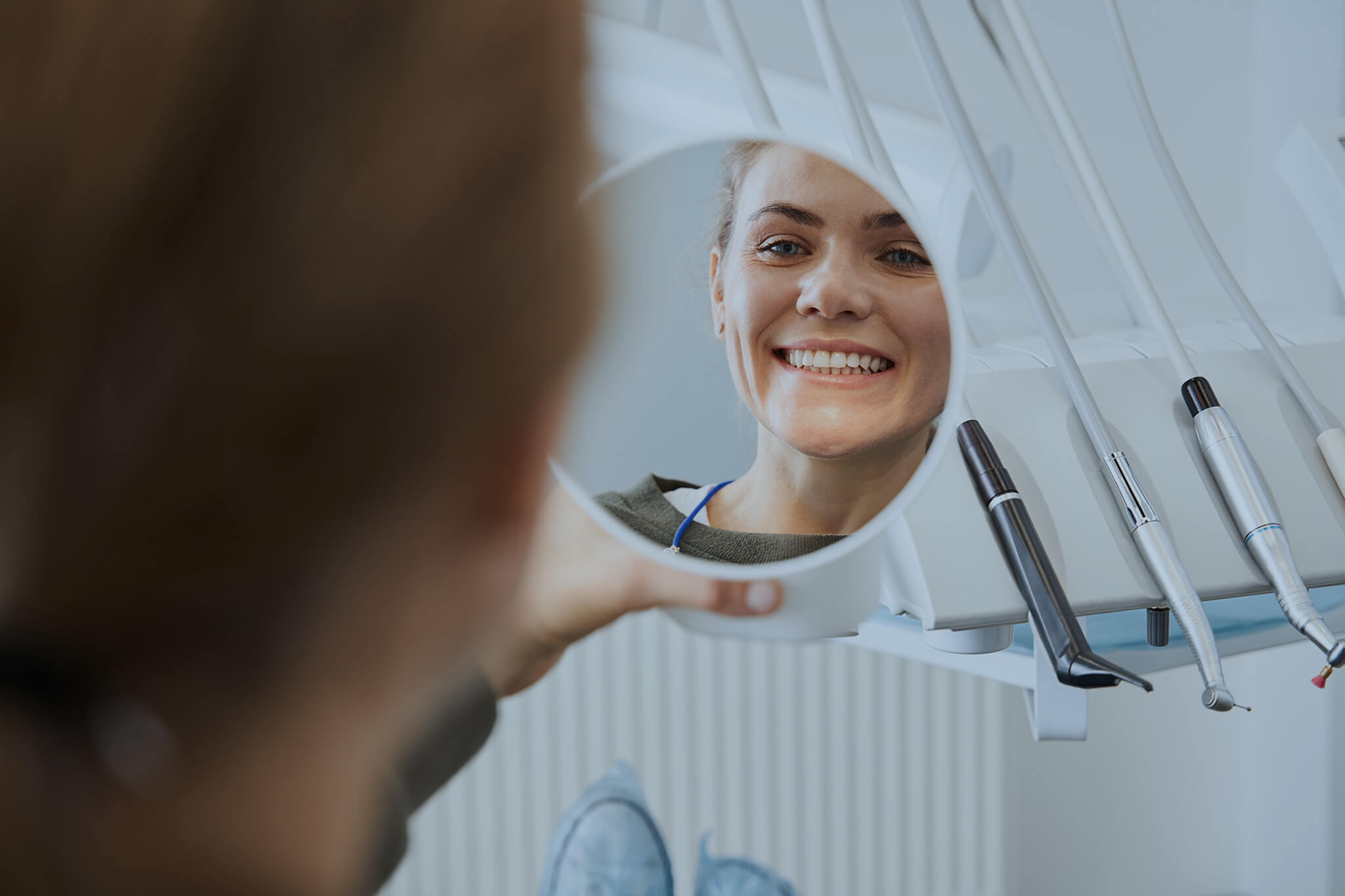
By selectively targeting soft tissue, gingivectomy with laser can yield a more precise and minimally invasive approach — often resulting in less bleeding, reduced discomfort, and faster healing.
In this article, we discuss the top reasons for performing a laser gingivectomy, explore the benefits of using laser technology, examine the types of lasers commonly employed, and provide a step-by-step guide on how to perform a laser gingivectomy effectively.
Reasons for Performing a Gingivectomy
Crown Preparation (Gingivectomy for Crown Prep)
The most common use for laser gingivectomy is crown preparation. Excess gum tissue can obstruct proper access to the tooth when preparing for a crown.
A crown prep procedure often requires removing a small amount of gingiva, and using a laser can help refine this process by offering a precise cut with minimal bleeding.

Aesthetic Improvement
Some patients have what’s referred to as a gummy smile, where the gum line appears disproportionately high or uneven.
Performing a gingivectomy with laser can remove and reshape the excess tissue, giving the patient a more balanced and attractive smile.
Periodontal Therapy
When gingival overgrowth or infected gum tissue impedes proper oral hygiene, a gingivectomy may be performed to reduce pockets and facilitate better brushing and flossing.
Laser-assisted techniques can be particularly beneficial in areas prone to bleeding or infection.
Pseudopocket Removal
Certain types of gingival overgrowth create false pockets that trap debris and bacteria.
By removing or reducing these pockets through laser gingivectomy, patients can maintain healthier gums, potentially avoiding more significant periodontal issues in the future.
Benefits of Laser-Assisted Gingivectomy
Traditional operculum dental treatments involve scalpels or electrosurgical units. While effective, these methods may result in bleeding, a higher risk of infection, and prolonged recovery times.
A laser-assisted operculectomy offers several advantages:
- Minimal Bleeding: Lasers cauterize blood vessels as they cut, reducing the need for sutures and decreasing postoperative bleeding.
- Sterilizing Effect: The heat from the laser helps disinfect the area, lowering the risk of bacterial contamination and post-operative infection.
- Patient Comfort: Many patients report less pain and anxiety with laser treatments than with traditional incisions, potentially reducing the need for anesthesia.
- Faster Recovery: By minimizing trauma to surrounding tissues, laser-assisted gingivectomy procedures often allow for a quicker return to normal eating and hygiene habits.
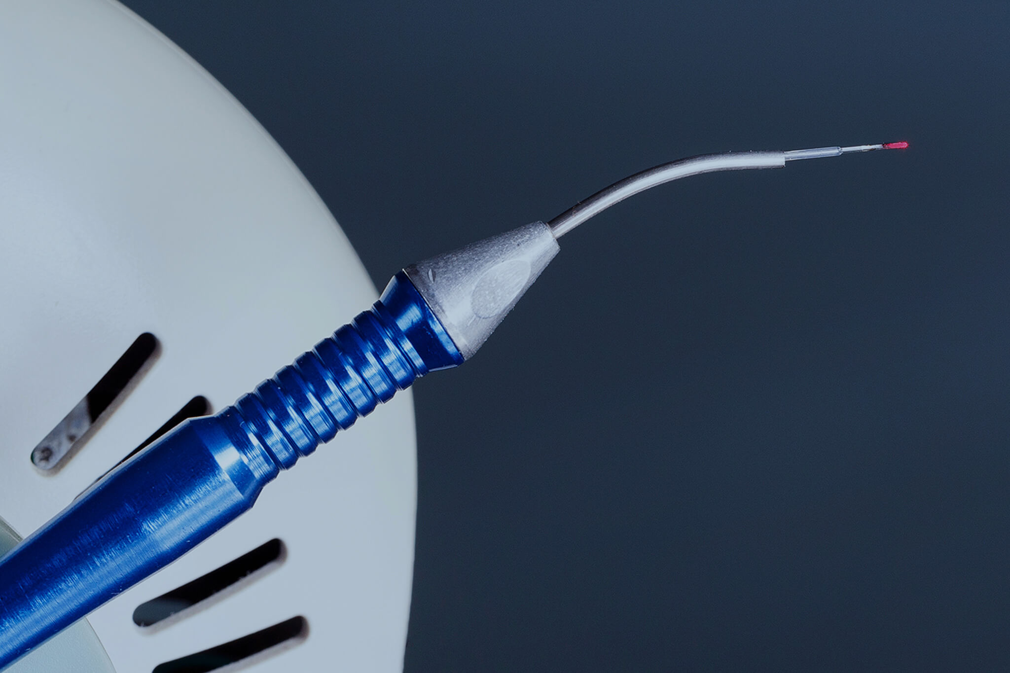
Types of Lasers Used in Gingivectomy Treatments
Different laser technologies can be selected based on the degree of soft-tissue removal, desired hemostasis, and the patient’s oral condition. The most common lasers used in operculectomy procedures include:
CO₂
CO₂ lasers are known for precise soft-tissue cutting and coagulation. Their wavelength is strongly absorbed by water-rich tissues, enabling accurate ablation with minimal thermal damage.
Diode
Compact and cost-effective, diode lasers work well on soft tissues containing hemoglobin or melanin. They’re frequently used for soft-tissue procedures like gingivectomies, given their reliability in controlling bleeding.
Nd:YAG
Nd:YAG lasers penetrate tissues more deeply, aiding in subgingival bacterial reduction — beneficial in cases where periodontal pockets or infected tissues are present.
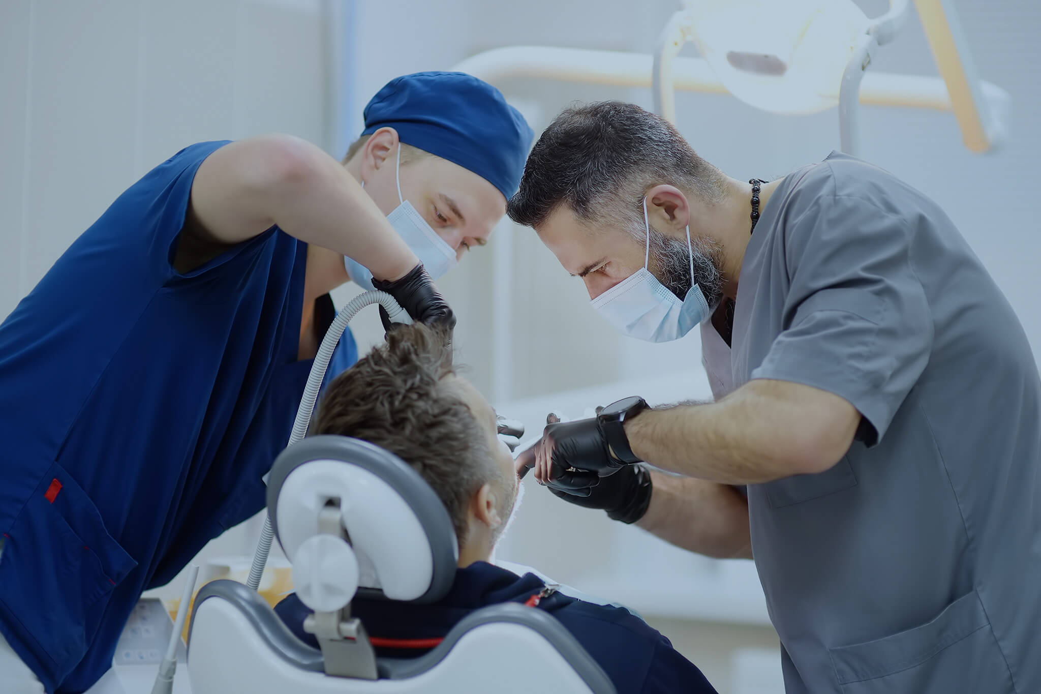
How Dental Surgeons Can Perform a Laser Gingivectomy Procedure
Optimize patient comfort, reduce bleeding, and achieve a more seamless transition between treated and healthy tissues by following these steps:
1. Diagnosis and Preparation
Evaluate the patient’s gum health and determine the exact amount of gingival tissue to remove.
Use radiographs or probing measurements to confirm that underlying bone support is adequate for reshaping.
Administer local anesthesia to ensure comfort and place protective barriers (such as a wax spatula) between the tooth and gum if needed, especially if the laser might come close to enamel or restorative materials.
2. Laser Selection and Calibration
Choose the appropriate laser type based on the thickness of gum tissue, patient sensitivity, and presence of infection or bleeding.
Calibrate power, pulse duration, and frequency to achieve precise cuts with minimal thermal damage.
3. Tissue Removal
Position the laser handpiece perpendicular to the gingiva, removing tissue in small, controlled passes.
Defocus the laser (by increasing the nozzle-to-tissue distance) if bleeding occurs, leveraging the device’s hemostatic properties and smooth the edges to ensure a natural transition between treated and healthy tissues.
4. Final Checks
Gently irrigate the area to remove debris and check for any residual pockets of inflamed tissue.
Confirm hemostasis — often, no sutures are needed if the laser has adequately sealed blood vessels.
5. Postoperative Care
Provide patients with home care instructions, including gentle mouth rinses, avoiding chewy or hard foods, and using prescribed anti-inflammatory or antibiotic medications if indicated.
Schedule follow-up visits to monitor healing and confirm that the gums have taken on the desired shape.
Partner with IML for Advanced Surgical Lasers
Although International Medical Lasers (IML) does not currently provide dental laser devices specifically for operculectomy, we do offer a diverse range of cutting-edge laser systems tailored to other specialties such as urology, gastroenterology, and ENT.
If you’re interested in expanding your practice’s laser capabilities beyond dental treatments, get in touch with IML to explore our portfolio of high-quality medical laser systems.