Operculectomy Dental Procedure with Laser Technology
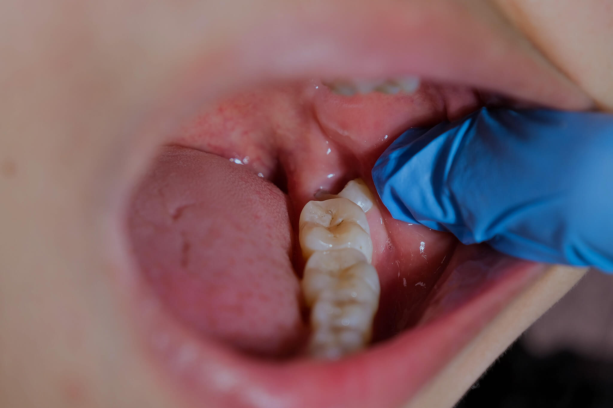
When left untreated, an operculum can trap food and bacteria, leading to discomfort, inflammation, and even infection.
In this article, we explore laser-assisted operculectomy techniques, the types of lasers used in operculum dental treatments, and the steps dental professionals can take to perform this procedure using modern, minimally invasive methods.

Benefits of Laser-Assisted Operculectomy Procedures
Traditional operculum dental treatments involve scalpels or electrosurgical units. While effective, these methods may result in bleeding, a higher risk of infection, and prolonged recovery times.
A laser-assisted operculectomy offers several advantages:
- Minimal Bleeding: Lasers cauterize blood vessels as they cut, reducing the need for sutures and decreasing postoperative bleeding.
- Sterilizing Effect: The heat from the laser helps disinfect the area, lowering the risk of bacterial contamination.
- Patient Comfort: Many patients report less pain and anxiety with laser treatments than with traditional incisions, potentially reducing the need for anesthesia.
- Faster Recovery: By minimizing trauma to surrounding tissues, laser-assisted procedures often allow for a quicker return to normal eating and hygiene habits.
Types of Lasers Used in Operculum Dental Treatments
Different laser technologies can be selected based on the degree of soft-tissue removal, desired hemostasis, and the patient’s oral condition. The most common lasers used in operculectomy procedures include:
CO₂
CO₂ lasers are known for precise soft-tissue cutting and coagulation. Their wavelength is strongly absorbed by water-rich tissues, enabling accurate ablation with minimal thermal damage.
Diode
Compact and cost-effective, diode lasers work very well on soft tissues containing hemoglobin or melanin, making them suitable for controlling bleeding.
Nd:YAG
Nd:YAG lasers penetrate tissues more deeply, aiding in subgingival bacterial reduction—this is important if pericoronitis has developed.
Erbium
Erbium lasers are useful for both hard and soft tissue. While typically known for cavity preparation or bone recontouring, erbium lasers can also address an operculum if minor bone trimming is needed.
How Dentists Can Perform a Laser Operculectomy
Optimize patient comfort, reduce bleeding, and achieve a more seamless transition between treated and healthy tissues by following these steps:
1. Diagnosis and Preparation
Begin with a thorough evaluation — often including radiographs — to confirm that the operculum is causing issues such as inflammation, infection, or occlusal trauma.
Although laser treatments may reduce the need for high-dose anesthesia, most clinicians administer a local anesthetic to ensure patient comfort.
If the operculum covers part of the tooth, consider placing an insulating barrier (like a wax spatula) between the tissue and the enamel to prevent accidental laser contact.
2. Laser Selection and Calibration
Select a suitable laser (CO₂, diode, Nd:YAG, or erbium) based on the tissue’s thickness, bleeding tendencies, and level of infection.
Adjust the laser settings — power, pulse duration, and frequency — to achieve precise ablation with minimal thermal damage.
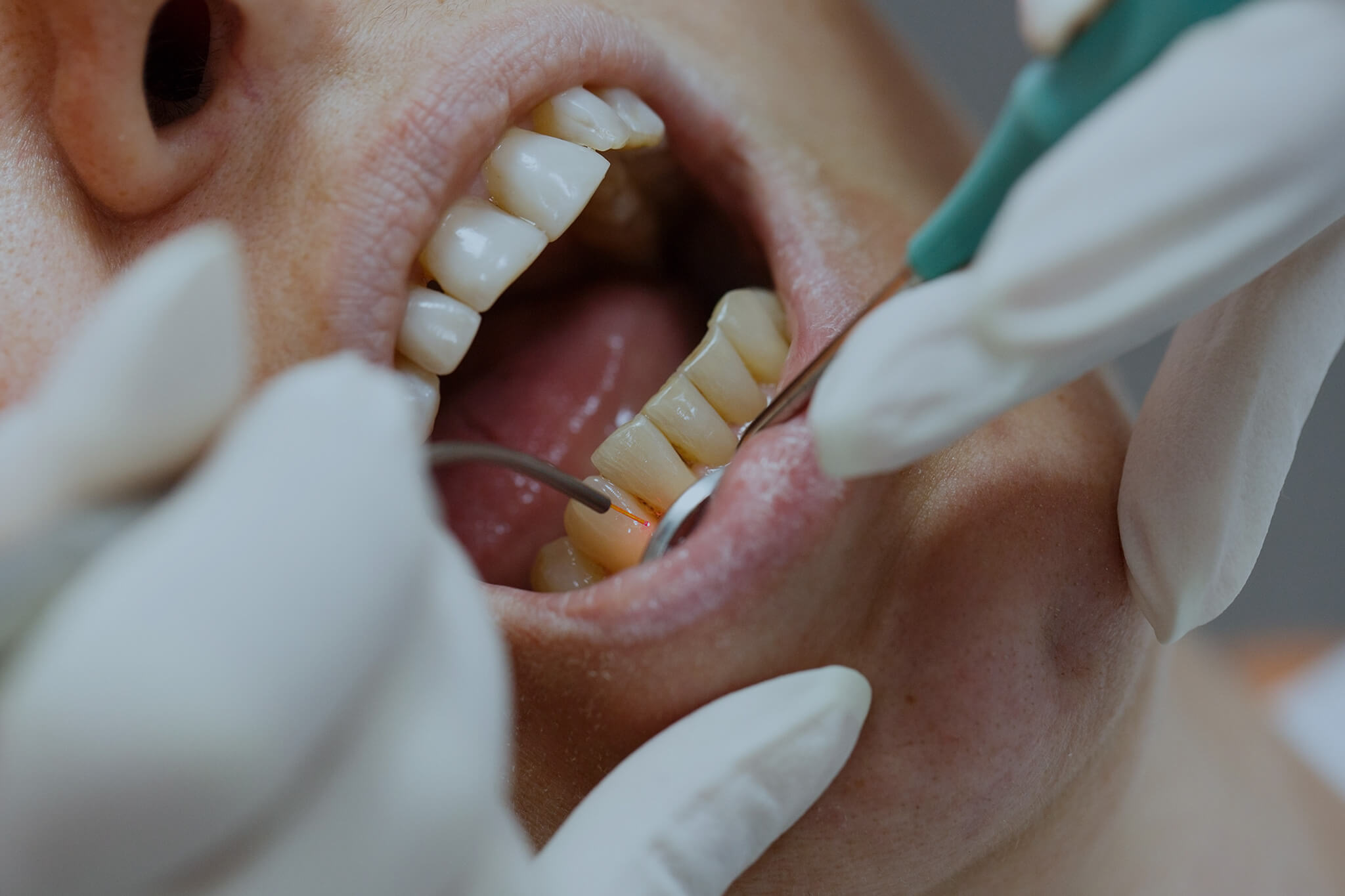
3. Tissue Removal
Carefully ablate the operculum in layers, positioning the laser handpiece perpendicular to the gum flap. As the laser vaporizes unwanted tissue, it also sterilizes the surgical field.
Defocusing the laser (by increasing the handpiece’s distance) can aid in achieving swift hemostasis when bleeding occurs.
4. Decontamination and Final Checks
After removing the main portion of the operculum, consider using a specialized laser tip (e.g., a periodontal tip) to decontaminate the sulcus, particularly if pericoronitis or bacterial infection is present.
Gently irrigate the site to clear any debris and confirm hemostasis, which often eliminates the need for sutures.
Lastly, verify that the remaining gum architecture is stable and free from residual tissue tags.
5. Postoperative Care
Provide patients with home care instructions, including gentle mouth rinses, avoiding chewy or hard foods, and using prescribed anti-inflammatory or antibiotic medications if indicated.
Follow-up appointments help assess healing progress, confirm resolution of pericoronitis symptoms, and ensure that no regrowth of the operculum is occurring.
Partner with IML for Advanced Surgical Lasers
Although International Medical Lasers (IML) does not currently provide dental laser devices specifically for operculectomy, we do offer a diverse range of cutting-edge laser systems tailored to other specialties such as urology, gastroenterology, and ENT.
If you’re interested in expanding your practice’s laser capabilities beyond dental treatments, get in touch with IML to explore our portfolio of high-quality medical laser systems.