Sclerotherapy Endoscopy Treatment
Adding a low-power endoscopic laser has refined that technique, giving endoscopists an immediate way to seal needle tracks, stabilize clots, and treat residual feeders through endoscopic injection sclerotherapy without changing scopes.
In this post, we’ll examine how laser-assisted sclerotherapy endoscopy works, which clinical problems benefit most, the step-by-step technique, and the laser platforms now making dual-modality therapy practical in everyday GI practice.

The Evolution of Sclerotherapy
Sclerotherapy first slipped into the endoscopy suite in the late 1960s, laying the groundwork for modern sclerotherapy endoscopy treatment. Surgeons used the same sclerosants they had been injecting into varicose veins and portal collaterals during open procedures.
Back then, quinine and sodium morrhuate were blunt instruments—they provoked fibrosis, but the price was deep ulceration and post-treatment strictures.
By the early 1980s, safer detergents such as ethanolamine oleate and sodium tetradecyl sulfate had replaced the harsher agents. Their surfactant action damaged the endothelium without scorching the surrounding mucosa, so bleeding control improved and complications fell.
The 1990s saw the development of endoscopic band ligation, a mechanical solution that quickly became first-line for oesophageal varices. Yet operators soon realised no single modality could tame every bleed—large gastric varices, tortuous Dieulafoy lesions, and diffuse radiation proctitis still demanded something more adaptable.
Enter hybrid laser-assisted sclerotherapy.
By pairing a familiar chemical thrombogen with a pinpoint beam of light, today’s endoscopist can inject, seal, and spot-treat residual feeders through the same scope channel. It is the latest step in a half-century movement toward faster haemostasis, fewer sessions, and gentler tissue handling.
How Modern Sclerotherapy Works
An engorged varix is like an over-inflated bicycle tube—thin-walled, high-pressure, and ready to burst.
When a sclerosant is injected, it strips the inner lining (endothelium), exposes collagen, and sparks immediate clotting. Within seconds, platelets aggregate and a fibrin web begins to form.
The adjunct laser then plays two crucial roles:
- It heat-seals the tiny puncture channel, preventing back-bleed that could wash the fresh clot away.
- Its thermal energy shrinks the surrounding collagen, compressing the varix like a snug elastic band. That gentle squeeze lowers shear stress long enough for the fibrin matrix to cross-link and harden into a stable thrombus.
Chemical injury starts the process; optical energy helps it forge a durable seal—all without switching devices or losing sight of the bleeding point.
What is Laser-Assisted Sclerotherapy Endoscopy?
The procedure combines two energy sources delivered through the same working channel:
- Chemical sclerosant: 1-2 mL of ethanolamine, polidocanol, sodium tetradecyl sulfate, or cyanoacrylate, all delivered via standard endoscopic injection sclerotherapy technique.
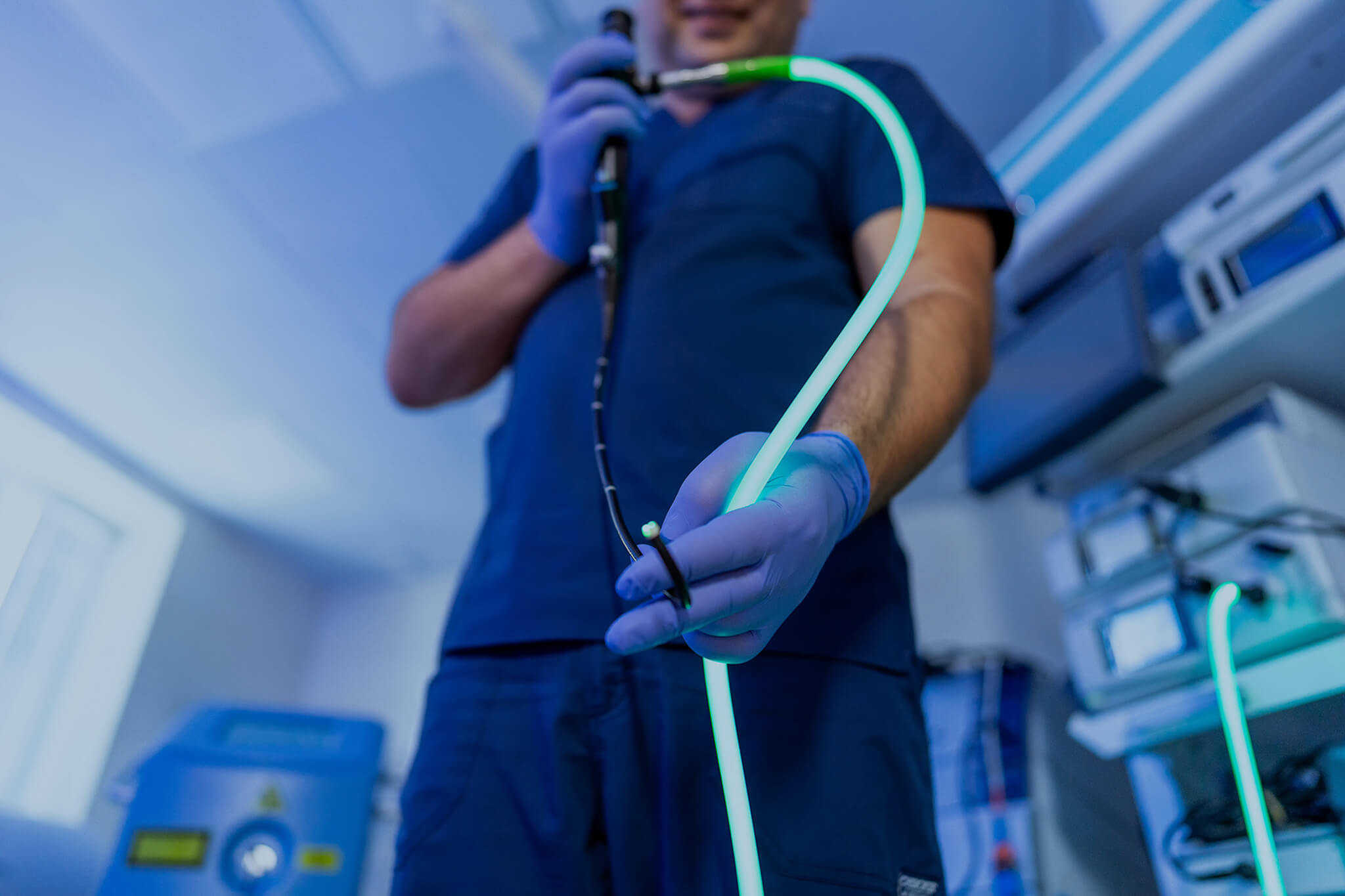
- Endoscopic laser light: short, 10-15 W pulses from a 600 µm diode or Nd:YAG fibre applied directly to the injection bleb.
Sclerosant initiates thrombosis, while laser pulses (usually 8-15 W for one second) photocoagulate the puncture site. Thermal energy denatures residual endothelium and shrinks surrounding tissue, reinforcing the chemical reaction.
Because both steps occur through a familiar accessory channel, teams already comfortable with standard injection adopt the technique quickly.
Together, chemical and optical energy shorten bleed-stop time, limit sclerosant back-bleed, and reduce the number of reinjection sessions.
Why Combine Laser and Sclerosant?
Traditional sclerotherapy endoscopy relies on chemistry alone. Most bleeds stop quickly, but a minority persist because injected columns leak or puncture holes reopen under portal pressure. The laser acts as a biological top-stitch, welding the entry site and coagulating surface vessels that escape the sclerosant halo.
Bench studies show that a one-second, 10-watt pulse seals a 21-gauge needle tract in porcine oesophagus in under three seconds. Clinical trials mirror those findings.
Benefits of Sclerotherapy Endoscopy
Randomised studies report acute control rates of 90-95% in oesophageal varices, with rebleed rates comparable to ligation when beta-blockade accompanies therapy. Practitioners cite several day-to-day benefits:
- Rapid haemostasis during sclerotherapy endoscopy procedures, even when visual fields are narrow.
- Accessible worldwide, as needles and sclerosant cost a fraction of ligation kits.
- Flexible dosing so the operator can treat multiple lesions in one session.
- Durable obliteration since fibrotic cords resist portal hypertension better than bands alone.
- Preserved mucosal contour for future dilations or stent placements.

Choosing the Right Sclerosant
Matching agent, concentration, and volume to vessel size improves efficacy. It also minimizes complications. Below is a list of sclerosants and their characteristics.
| Agent | Typical concentration | Preferred lesions | Key strengths | Important caveats |
|---|---|---|---|---|
|
Ethanolamine oleate 5 % |
1-2 mL blebs |
Oesophageal varices |
|
Mild chest pain; transient fever |
|
Sodium tetradecyl sulfate 1-3 % |
0.5-1 mL blebs |
Small varices Haemorrhoids |
|
Tissue necrosis if extravasated |
|
Polidocanol 1-2 % |
1-2 mL lines |
Radiation proctitis AVMs |
|
Multiple sessions often required |
|
N-butyl-2-cyanoacrylate (0.5 mL) + lipiodol |
Varices ≥10 mm |
Gastric varices |
|
Embolism risk Inject ≤1 mL per shot |
Laser Platforms Suited to the Practice
Most units already house soft-tissue lasers for Barrett’s ablation or airway surgery. The same consoles adapt easily to GI haemostasis when paired with sterilizable 600 µm fibres that pass through 2.8 mm channels.
| Platform | Wavelength | Power range | Notable GI features |
|---|---|---|---|
|
1064 nm |
5-20 W |
|
|
|
1940 nm |
2-6 W |
|
All accept autoclavable 600 µm GI fibres that pass through standard 2.8 mm channels, so no specialty scope is required. Each platform offers foot-pedal control, standby safety interlocks, and integration with existing smoke evacuation.
When to Consider Laser Assistance in Sclerotherapy Endoscopy Treatments
Hybrid therapy is most useful when anatomy or clinical urgency limits band ligation, over-the-scope clipping, or pure chemical injection.
Before detailing indications, clinicians should remember that laser fibres deliver energy forward, so clear visual fields remain essential.
Typical scenarios include:
- Acute oesophageal variceal bleeding with active spurting.
- Fundal or isolated gastric varices deeper than 10 mm below the mucosa.
- Large-calibre Dieulafoy lesions that re-bleed after epinephrine alone.
- Diffuse radiation proctitis where telangiectasia carpet wide areas.
- High-flow internal haemorrhoids in patients on anticoagulation.
Patient Selection and Safety
Most candidates mirror those for conventional endoscopic injection sclerotherapy, but the hybrid approach is advantageous when small, tortuous vessels make band capture unreliable.
Absolute contraindications remain uncorrected coagulopathy, allergy to the planned sclerosant, and airway instability that prevents safe endoscopy during active haemorrhage.
Relative cautions include prior extensive banding scars that narrow the lumen, recent oesophageal dilation, and known strictures that could impair fibre manoeuvrability.

Pre-Procedure Preparation
Endoscopy nurses confirm NPO status, check blood counts, and start proton-pump inhibitors or vasoactive agents when cirrhosis is present. Massive bleeds may need intubation before the procedure begins.
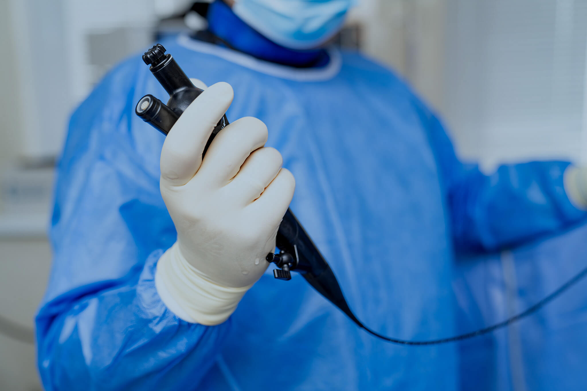
A 21 G injection needle, sclerosant vial, sterile 600 µm fibre, and laser console foot-switch are placed on the procedural cart.
Step-by-Step Laser-Assisted Sclerotherapy Endoscopy
The hybrid sclerotherapy endoscopy procedure sequence adds less than a minute to standard injection times. Most cases proceed under moderate sedation, using midazolam and fentanyl titrated to patient comfort. A typical workflow includes:
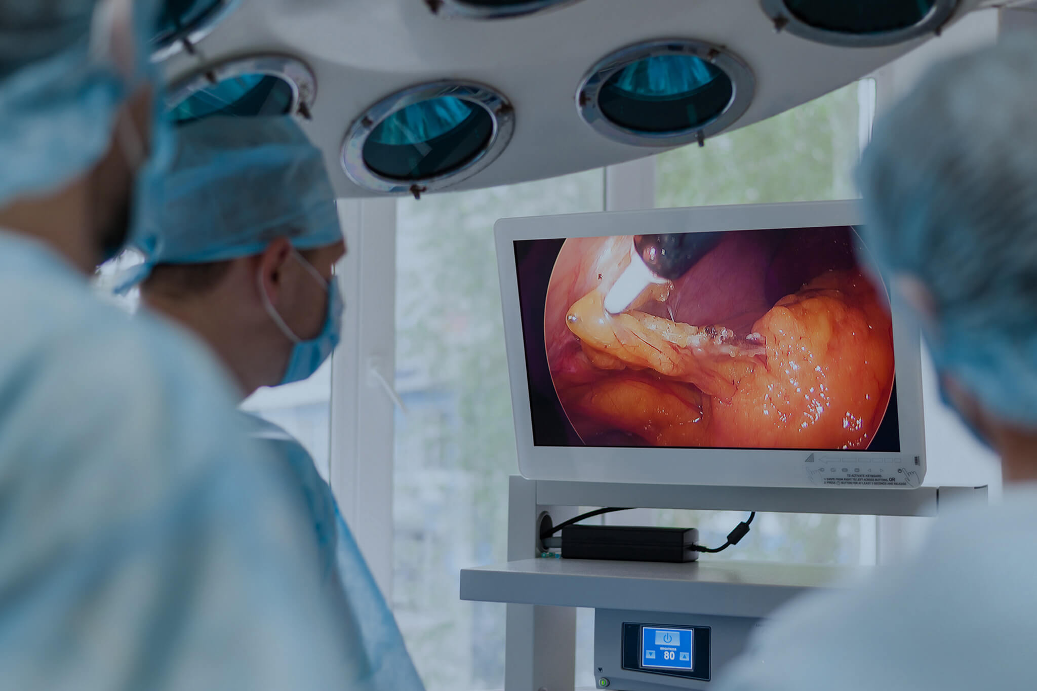
- Identify the bleeding point and suction pooled blood to clear the field.
- Prime the needle with sclerosant to avoid dilution.
- Inject 0.5–2 mL directly into the varices until blanching or distention is observed.
- Withdraw the needle and introduce a sterilised fibre without removing the scope.
- Deliver a 10W, one-second pulse 1 mm off the bleb apex, producing a tan seal.
- Apply additional 0.5-second pulses to residual feeders or oozing points.
- Flush the fibre tip with saline, document sclerosant volume and total joules, and proceed to the next lesion.
Recovery and Aftercare
Successful hemostasis is only the first step. Structured follow-up keeps patients comfortable and catches early recurrences. The post-procedure plan is simple but disciplined, moving from diet advancement to pharmacologic protection and scheduled surveillan
Diet Progression
- Clear liquids for the first four hours
- Transition to a soft diet as tolerated and avoid rough or spicy foods for 48 hours
Pharmacologic Protection
- Continue a proton-pump inhibitor twice daily
- Add sucralfate suspension four times daily for seven days to minimise ulcer risk

Early Surveillance
- Perform an endoscopy on day 7 to confirm thrombosis
- Treat any residual varices with band ligation or a second hybrid injection during the same session
Long-Term Monitoring
- Schedule quarterly surveillance scopes until portal pressure is definitively lowered—typically after a TIPS procedure or liver transplantation
- Reevaluate beta-blocker therapy and adjust based on hemodynamics and endoscopic findings
Complications and Risk Mitigation
Complication patterns resemble those of traditional sclerotherapy but occur less often when laser settings remain conservative.
| Event | Mitigation strategy |
|---|---|
|
Superficial ulceration |
|
|
Oesophageal stricture |
|
|
Deep wall necrosis |
|
|
Cyanoacrylate embolism |
|
|
Post-procedure pain |
|
Training Requirements and Learning Curve
Endoscopists comfortable with standard injection adapt rapidly, but a few new competencies ensure safety and efficacy. Consider the following training components.
Laser-Safety Protocols
- Complete formal laser-safety certification covering:
- Eye protection (e.g., ANSI-rated goggles).
- Console foot-pedal operation and standby interlocks.
- Smoke-plume management via dedicated evacuation ports.
- Understand laser-tissue interactions—thermal penetration depth at 10-15 W pulses—to avoid deep wall necrosis.
Fibre Handling and Preparation
- Inspect each 600 µm fibre tip under magnification for cracks or debris.
- Condition the fibre by priming with saline to remove air bubbles.
- Maintain a 1-2 mm gap between fibre tip and mucosa—too close can perforate; too far reduces coagulation efficiency.
- Retract the fibre slowly to prevent kinking and preserve the quartz core.
Foot-Pedal Coordination
- Begin with hands-off practice on ex vivo models (animal tissue or synthetic simulators).
- Train to deliver a one-second pulse only when the fibre is stationary above the bleb.
- Nurse-led “timeout” protocols ensure console settings (pulse width, power level) match the lesion type.
Integration with Injection Technique
- Because most endoscopists already place 21 G needles through a 2.8 mm channel, adding a 600 µm fibre is intuitive.
- Emphasize seamless scope-insertion steps:
- Inject until the varix blanches or bulges (0.5-2 mL).
- Withdraw the needle and immediately insert the conditioned fibre without scope removal.
Mentored Proctoring
- Schedule the first 3-5 hybrid cases under an experienced laser-assisted sclerotherapy mentor.
- Mentor guides:
- Ideal pulse-power adjustments for varying bleb sizes.
- Troubleshooting visual challenges, such as smoke cloud buildup.
- When to irrigate with saline to clear charred debris.
Ongoing Skill Refinement
- Maintain a simple laser-usage log tracking:
- Total joules delivered per case.
- Any adverse events or unexpected tissue reactions.
- Review logs monthly to optimize:
- Power settings for common lesion types (e.g., large gastric varices vs. telangiectasia).
- Fibre re-use guidelines per manufacturer to balance safety and cost.
Implementing these training steps—eye protection, fibre prep, foot-pedal drills, proctored cases, and log reviews—shortens the learning curve. Within 3-5 hybrid procedures, an experienced injector can deliver laser-assisted sclerotherapy as efficiently as pure injection alone.
Key Takeaway for Surgical Teams and Device Buyers
Laser-assisted sclerotherapy endoscopy blends the deep thrombogenic effect of chemical injection with the immediate surface seal of photocoagulation.
Units already equipped with soft-tissue lasers can adopt this hybrid method quickly, improving hemostasis, lowering re-bleed rates, and reducing total sclerosant exposure without major capital investment.
For GI services seeking a nimble, cost-effective upgrade that complements band ligation and TIPS, hybrid sclerotherapy is an evidence-based option ready for immediate deployment.
Get in touch with Us
If you have questions about which laser is right for your practice, speak with our expert team today.